Campagna 2024

em web banner 1200x480

Download, read and share.

Ā 

Feel free to download our educational material and share them with your family, friends, colleagues...

Ā 

A short film from Euromelanoma about life after skin cancer diagnosis

A short film from Euromelanoma about life after skin cancer diagnosis (with English subtitles)

Leaflet

Poster

Skin Check Guide

Patient Charter

Digital poster - 5s

Ā 

Digital poster - 10s

Ā 

Preventing skin cancer

Skin cancer is one of the worldā€™s most common cancers. The good news is that it is usually treatable if detected early - and there are things we can do to prevent it.

Always be sunsafe

Avoid unnecessary exposure.

Seek shade where possible, and avoid the summer sun during the middle of the day.

sun icon

Wear protective clothing

Include dark colours, long sleeves, a widebrimmed hat and UV-rated sunglasses.

For children, look for clothing with inbuilt sun protection.

clothes icon

Apply sunscreen

Check that yours has a high protection factor against both UVA and UVB rays. Remember that sunscreen takes effect around half an hour after its application and only lasts for two to three hours.

uv icon

Protect children

Children are at the greatest risk of long-term health issues related to unsafe sun exposure.

Outside play is important, but you should never let a child get sunburnt.

baby icon

What to look for

Ā 

Regularly examining your skin is important for catching potential signs of skin cancer early. Here are some key indicators to watch out for:Ā 

Ā 

  • New moles: Keep an eye out for the appearance of new moles, particularly in adulthood. Ā 
  • Changes in size, colour, and shape: Any spot on your skin that undergoes changes in size, colour, or shape deserves attention.Ā 
  • The 'Ugly Duckling' sign: If you notice a mole that stands out from the rest, it should be examined by a dermatologist.
  • Asymmetry or uneven borders: Normal moles are mostly symmetrical and have even borders. If you notice irregularities in symmetry or borders, have it checked out.
  • Multicolour: Keep an eye out for spots with multiple colours or shades (black, brown, tan, white, grey, red, pink, or blue).
  • Texture: Pay attention to how skin lesions feel. Rough or scaly lesions (e.g. on the face) may be detectable by touch before they become visible.
  • Non-healing wounds: Wounds or sores that don't seem to heal, or heal very slowly, might be something other than an injury.
  • Bleeding, oozing or itching: Spots that bleed without apparent cause, ooze or itch should be examined.Ā 
  • Pearly appearance: A shiny or pearly appearance can be indicative of skin cancer.

Ā 

If you notice one of these warning signs, it's crucial not to delay. Schedule an appointment with your dermatologist or healthcare provider for an examination. Early detection increases the chances of successful treatment.Ā 

Melanoma

Melanoma originates from melanocytes, the pigment-producing cells in the skin. It is less common than other skin cancers but is the most concerning one due to its potential for rapid spread and metastasis. Unlike other types that often affect older individuals, melanoma can develop in individuals of any age. It typically manifests as a mole or spot on the skin that undergoes noticeable changes over time. These changes may include darkening in color, irregularities in shape or borders, and the development of various hues within the lesion. Additionally, melanoma may present as a rapidly growing pink or red lump While it can arise from a common mole or dysplastic nevus, the majority develops in an area of seemingly normal skin. Melanomas can develop in other parts of the body than the skin, including inside the eye (ocular melanomas) or in the nose, mouth, throat, genital region, or anal area (mucosal melanomas). Itā€™s crucial to seek immediate medical attention if you notice any of these changes, as melanoma can spread internally and become life-threatening if left untreated.Ā 

Basal cell carcinoma

Basal cell carcinoma originates from the basal cells of the skin, located in the lowest layer of the epidermis. It is the most common type of skin cancer, yet it tends to be less aggressive compared to melanoma. Prolonged sun exposure is the primary cause, with basal cell carcinoma typically affecting sun-exposed areas. The presentation varies from a raised, skin-coloured bump with a smooth, shiny surface reminiscent of a pearl, to a non-healing sore, a reddish patch, or a scar-like area. Occasionally, it may manifest as a slow-growing bump with a slightly crusty texture at the center. While basal cell carcinomas grow slowly and rarely metastasize, prompt treatment is necessary to prevent further tissue invasion and damage.Ā 

Squamous cell carcinoma

Squamous cell carcinoma originates from the squamous cells of the skin. It ranks as the second most prevalent type of skin cancer, typically developing in regions of the skin extensively exposed to sunlight, such as the face and scalp. It may occur in other locations, like the genital area. Squamous cell carcinoma often appears as a crusty lump that may exhibit rapid growth and subsequently become ulcerated and exude fluid. Furthermore, it can present as reddish patches, open sores, scar-like areas, or warty lesions This cancer has the potential to spread swiftly, particularly when located on sensitive areas like the lips, ears, fingers, and toes, or in individuals with weakened immune systems, such as organ transplant recipients. Surgical intervention to excise the lesions is imperative for effective treatment.Ā 

Actinic keratosis

Actinic keratosis is a common precancerous skin condition resulting from cumulative sun exposure, predominantly affecting individuals in their middle-aged to elderly years. These lesions typically manifest as red-brown, scaly patches on areas of the skin exposed to sunlight, such as the face, scalp, ears, and hands. Approximately 10-15% of actinic keratoses may progress to squamous cell carcinoma if left untreated. Thus, prompt intervention through various treatment options is imperative to halt the potential progression to invasive skin cancer. Treatment modalities include topical medications, cryotherapy (freezing), or photodynamic therapy, depending on the severity and extent of the lesions. Early diagnosis and management play a crucial role in reducing the risk of developing more advanced skin cancers.Ā 

Merkel cell carcinoma

Merkel cell carcinoma, originating from specialized neuroendocrine Merkel cells in the skin's basal layer, is a rare yet aggressive skin cancer. With an incidence rate of about 0.59 cases per 100,000 individuals in Europe, it predominantly affects those over the age of 50. Typically asymptomatic, these rapid growing, reddish-yellowish spherical-looking tumours preferentially grow on chronically sun-exposed skin areas of the face and extremities. The primary causative factors are Merkel cell polyomavirus (MCPyV) infection and UV radiation exposure. Early detection and treatment, often involving surgery, radiation therapy, and immunotherapy, are crucial due to its aggressive nature and potential for metastasis.Ā 

Melanoma can be life-threatening if not detected and treated early. Therefore, it's essential to be familiar with the signs to look for. The ABCDE method is a useful tool for detecting melanoma:Ā 

Ā 

Ā 

Ā 

Ā 

A. Asymmetry: One half of the spot does not match the other half.Ā 

Ā 

Benign

Ā 

Malignant

B. Border irregularity: The edges of the spot are not smooth and may be uneven or blurred.Ā 

Ā 

Benign

Ā 

Malignant

C. Colour variation: The spot contains different colours or shades, such as brown, black, red, white, or blue.Ā 

Ā 

Benign

Ā 

Malignant

D. Melanomas are often larger in diameter than a pencil eraser (6mm).Ā 

Ā 

Benign

Ā 

Malignant

E. Evolution: The spot changes over time, growing larger, changing colour, or becoming raised.Ā 

Ā 

Before

Ā 

After

By paying attention to these ABCDEs, you can better identify potentially concerning spots on your skin and seek medical attention if needed. Remember, early detection is key to successful treatment and improved outcomes for melanoma.Ā 

Ensure you examine your entire body, including front and back, paying close attention to areas exposed to the sun. Use a combination of a full-length mirror and a hand mirror to thoroughly inspect all areas, including those difficult-to-reach spots.Ā 

1. Examine your face, including your nose, lips, mouth, and the areas behind and around your ears.Ā 

how 1

2. Check your scalp, using a comb to part your hair for better visibility during the examination. If you do not have much hair, it is especially important to thoroughly inspect your entire scalp.Ā 

how 2

3. Check the front and back of your hands and in between your fingers.Ā 

how 3

4. Then focus on your neck, chest, and upper body. Ensure you check between and underneath your breasts if applicable.Ā 

how 4

5. Bend your elbow to check your upper arm and armpits.

how 5

6. Use your hand mirror to check your neck and your back.Ā 

how 6

7. Check your buttocks and the back of your legs. Finish by checking the soles of your feet and in between your toes.Ā 

how 7

Skin cancer can affect individuals of any age, but it is most prevalent among those over 50 years old or those with prolonged sun exposure. One is at an increased risk if they:Ā 

Ā 

  • Have fair skin or are prone to sunburn
  • Have fair skin, light hair or eye colourĀ 
  • Have frecklesĀ 
  • Have more than 50 molesĀ 
  • Have a personal or family history of skin cancerĀ 
  • Experienced sunburn during childhoodĀ 
  • Spent significant time in the sun for work or leisure activities (chronic sun exposure)Ā 
  • Undergo periodical sun exposure, such as during holidays (intermittent sun exposure)Ā 
  • Use or ever have used sunbeds (particularly at a young age)Ā 
  • Are immunosuppressed (Organ transplant recipients, autoimmune diseases, HIV, drugs)Ā 
  • Are over the age of 50Ā 

Ā 

Regardless of your risk level, there are proactive measures you can take to protect yourself and your loved ones from skin cancer. By safeguarding your skin, performing regular self-checks, and being aware of warning signs, you can intercept any suspicious skin lesions before they escalate into more serious issues.Ā 

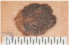
Skin cancer treatment options are available, and early detection significantly increases the likelihood of a successful recovery. If you notice a suspicious lesion, it is crucial to have it promptly evaluated by a doctor, preferably a dermatologist. Delaying treatment can exacerbate the condition, potentially leading to disfigurement, complications, or even death. Donā€™t let delay reduce your chances of a positive outcome.Ā 

Ā 

Follow these golden rules:

Ā 

  • Act instead of ignoring it, hoping it will resolve on its own
  • Avoid waiting to see how it progresses or attempting self-management
  • Refrain from assuming it's trivial or not urgent
  • Recognize the importance of addressing it promptly
  • Above all, don't hesitate to seek medical advice from your doctor or dermatologist

Ā 

Remember, skin cancer is manageable when detected early. If you notice any suspicious spots, make an appointment to see your doctor without delay.Ā